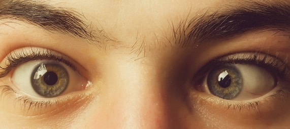
WHAT IS STRABISMUS AND LAZY EYE?

Strabismus is a visible misalignment of the eyes, with one eye turning in, out, up, down, or oblique.

Six eye muscles, controlling eye movement, are attached to the outside of each eye. One muscle moves in the eye to the right in each eye, and one muscle moves the eye to the left. The other four muscles move it up or down and at an angle.

How is strabismus treated?
Treatment aims to improve the alignment of the eyes and to bring back or protect normal vision. Treatment can include glasses, prisms, patching or blurring of one eye, eye drops, eye muscle surgery or botulinum toxin, which can alter eye muscles and eye exercises.
Blepharoplasty surgery repairs droopy eyelids that sometimes can cause blurred vision in one eye or both eyes by removing excess skin, muscle and fat.
It may be done for aesthetic purposes or where fields of vision are reduced.
Most strabismus is concomitant. Concomitant means the squint is the same in all angles of gaze. A variety of treatments are available for this to get rid of diplopia and improve the cosmesis. The options include surgery, botulinum injections, exercises, refractive or prism correction. Each case really needs to be judged on its own merits.
READ MORE ABOUT STRABISMUS
Strabismus misalignment of the eyes can occur in some or all directions of gaze and result in diplopia (double vision) or amblyopia (lazy eye).
Early diagnosis of strabismus is essential in preventing irreversible vision loss later in life. Strabismus treatment aims to improve the eyes’ alignment and correct the resulting vision loss (amblyopia). Amblyopia is regarded clinically as a 2-line difference from best-corrected visual acuity in a structurally healthy eye.
Strabismus is one of the most common eye conditions affecting up to 5% of the Australian population. Prisms are used to measure the degree of strabismus and double vision.
All of the muscles in each eye must be coordinated by the brain for binocular vision to function.
Complex neural networks link numerous structures. Interactions exist between these structures within each system separately, as well as between both systems. The ideal is for the best possible acuity of each eye, binocular (stereoscopic) vision, alignment of the eyes and accurate movements and saccades.
Strabismus may have sensory and/or motor origins from the peripheral and central nervous systems (brain and spinal cord ). Peripheral origin is at the level of the oculomotor muscles themselves or their innervation.
Often the squint or lazy eye at birth has its extraocular muscle imbalance associated with uncorrected hypermetropic refractive error.
In this type of strabismus, the eyes turn inward when the child focuses the eyes to see clearly. This crossing may occur when focusing at a distance, up close or both.
Such uncoordinated eye movements can induce vision loss as well as the loss of depth perception.
New strabismus in a school-aged child or adult necessitates further neurological investigations and medical histories such as malignancy or autoimmune conditions, exposures, vaccinations, fever, trauma, particularly to the head, orbit or periorbital area.
Signs such as gait disturbance, nystagmus, decreased visual acuity, or diplopia is relevant. The pattern of symptoms is essential, such as whether the strabismus is constant or intermittent. If intermittent, consider whether the symptoms have been changing in frequency and whether they are dependent on the direction of gaze or exaggerated by fatigue.
These muscle imbalances can be best diagnosed by the Hess Screen, the latest versions of which can be conducted by computer technology.
• Inward turning is called esotropia
• Outward turning is called exotropia
• Upward turning is called hypertropia
• Downward turning is called hypotropia.
Other classifications of strabismus include:
• The frequency with which it occurs—either constant or intermittent
• Whether it always involves the same eye—unilateral
• If the turning eye is sometimes the right eye and other times the left eye—alternating.
Typically, constant large-angle strabismus does not cause eye strain and headaches because there is virtually no attempt by the brain to straighten the eyes. Large-angle strabismus usually causes severe amblyopia in the turned eye if left untreated.
Whereas less noticeable cases of small-angle strabismus are more likely to cause disruptive visual symptoms, especially if the strabismus is intermittent or alternating.
In addition to headaches and eye strain, symptoms may include an inability to read comfortably, fatigue when reading and unstable or “jittery” vision. If small-angle strabismus is constant and unilateral, it can lead to significant amblyopia in the misaligned eye.
Ocular deviation may be manifest (tropia) or latent (phoria). The manifest ocular deviation can be present in all directions of gaze (comitant) or only present in specific directions of gaze (incomitant)
Concomitant deviations are relatively standard. They are usually associated with hypermetropia or the anomalous placement of one or more of the extraocular muscles.
Incomitant deviations are usually caused by a functional anomaly of one or more extraocular muscles or their associated neurology. This may result from a defective muscle or mechanical interference in the orbit (myogenic) or a consequence of a lesion in the nerves supplying the musculature (neurogenic).
The presence of diplopia usually suggests that the incomitancy is of recent origin and indicates a disturbance to some oculomotor system components. This may have been caused by trauma or may reveal the presence of an intracranial tumour, aneurysm or haemorrhage.
A detailed analysis of the oculomotor fields can be obtained using the Hess screen (Thomson Hess Pro on PC).
This is more effective and accurate than standard screening tests:
Light reflex test: The position of the light’s reflection in each of the eyes is compared. Deflection of the light reflex indicates abnormal ocular alignment, with each 1 mm of deflection equivalent to 15–20 prism diopters deviation.
The red reflex test: Comparison of the red reflex of both eyes simultaneously and noting asymmetries.
Cover and Uncover tests: Eye movements by the subject patient are compared.
However, the Hess results of the plots for the left and right eyes are the gold standard. They indicate which eye has a palsied muscle and provides an index for monitoring the progression of an incomitant deviation.
Motility is tested commonly in nine positions of gaze for each eye but can be done in 25 positions of gaze. The grid can be specified in units of degrees or prism dioptres. A prism dioptre corresponds to a deviation of 1 cm at 1 metre, which is equivalent to 0.573 degrees.
Primary causes of strabismus and secondary causes of strabismus can exist further to :
Congenital syndromes Cranial nerve palsies (CN III, IV, VI)
Orbital fracture
Intracranial bleed
Intracranial/intraorbital/intraocular mass (benign or malignant)
Intracranial infection
Grave’s disease
Myasthenia gravis
Diabetes mellitus
Toxins and heavy metal poisoning
Post-vaccination
Fevers
To line up and focus both eyes on a single target, all of the muscles in each eye must be balanced and working together. For the eyes to move together, the muscles in both eyes must be coordinated. The brain controls these muscles.
With normal vision, both eyes aim at the same spot. The brain then combines the two pictures into a single, three-dimensional image. This three-dimensional image gives us depth perception.
What are the symptoms of strabismus?
How is vision affected?
Good vision develops during childhood when both eyes have normal alignment. Strabismus may cause reduced vision, or amblyopia, in the misaligned eye.
Strabismic amblyopia occurs in approximately half of the children who have strabismus.
Amblyopia can be treated by patching or blurring the stronger eye to strengthen and improve vision in the weaker eye. If treatment is delayed, amblyopia may become permanent. The earlier amblyopia is treated, the better the result for vision. Family history can be relevant.
If one eye becomes favoured, it can become dominant and suppress the effect of the other eye vision integrating with the brain. To stop confusion, the brain will sometimes ignore part of the image from one eye.
What are the signs and symptoms?
Common signs can include closing one eye, clumsiness, an unusual head position, misaligned eyes, double or blurred vision and difficulty reading.
It is usual for a baby’s eyes to look misaligned for short periods up to four months, but full assessment is required.
Strabismus is especially common among children with disorders that may affect the brain, such as:
Cerebral palsy
Down syndrome
Hydrocephalus
Brain tumours
Prematurity
Genetics and family history are relevant. If you or your spouse has strabismus, your children have a greater risk of developing strabismus as well.
Some intermittent and small-angle strabismus cases may be possible to improve eye alignment non-surgically with vision therapy.
For example, convergence insufficiency (CI) is a specific type of intermittent exotropia. The eyes usually align properly when viewing a distant object but fail to achieve or maintain proper alignment when looking at close objects, such as when reading resulting in one eye drifting outward. Convergence insufficiency can interfere with comfortable reading, causing eye strain, blurred vision, double vision and headaches.
Children with exotropia (including convergence insufficiency) at an early age were significantly more likely to develop attention deficit hyperactivity disorder (ADHD), adjustment disorder and learning disabilities by early adulthood.
Certain types of ecotropic strabismus also have been associated with an increased risk of myopia, but vision therapy combined with eye exercises can help.
In children born with strabismus, it can be due to muscles or nerves not developing correctly. Still, it is usually due to the brain not controlling eye alignment accurately if it appears in the first few months of life. Children who have a problem with the focus of their eyes, especially those who are longsighted, can develop strabismus. Strabismus can also appear after head injuries or illnesses that cause weakness in eye muscles.
In strabismus surgery, one or more of the eye muscles are strengthened, weakened or moved to a different position by permanent or adjustable suture to improve alignment.
Patching or blurring the strong eye to improve amblyopia (development of lazy eye due to lack of stimulation) is often necessary. For accommodative esotropia, glasses reduce the focusing effort and often straighten the eyes. Sometimes bifocals are needed for close work.
Sometimes, the fold of skin at the inner eyelid can make eyes appear crossed with a wide, flat nose. This appearance of pseudostrabismus may improve as the child grows.
Adults who develop strabismus suddenly often have double vision because their brains have already learned to receive images from both eyes and cannot ignore the image from the turned eye. A child generally does not see double.
New incomitancies should get a medical workup, with or without diplopia.
For abnormal retinal correspondence (ARC), and suppression Bagolini lenses can be used. ( to determine when a line or part of the line disappears )
A Worth, 4 Dot test for peripheral sensory fusion and foveal suppression at different distances can be used. (Angular subtense gets larger at closer distances ).
Most strabismus with childhood deviations suppress and do not experience diplopia. Incomitant squints occur if there is an anomaly in the muscle or the nerves serving the muscles or some form of a space-occupying lesion. Noncomitant deviations that are new in adulthood generally cause diplopia, at least in some fields of gaze. Over time, a series of sensory and motor adaptations tend to occur, which reduce diplopia.
Suppression is one of the neural adaptations that can occur to reduce diplopia.
Suppression zones can be mapped by binocular perimetry or by prism measurements to induce double vision.
The Hess screen helps determine which muscle(s) are under and over-acting, and if a target wasn’t seen, evidence of suppression occurs.
In non-comitant cases, sometimes ‘sector’ prism is used, using Fresnel or press-on prism. The prism can be cut and adheres to the glasses, so it can be placed in the direction that the person needs it (cosmesis is considered inferior, and it reduces contrast sensitivity, though). Another method is to use sector occlusion, where a Bangerter filter or similar translucent, press-on filter blocks the vision in a particular field of gaze.
A Binocular Vision Analyser contains a Hess Screen and Field of Binocular Single Vision test. The former, of course, measures dissociated angle of deviation in each direction of gaze. The latter measures area of the field where the patient experiences single and double vision.
A new incomitancy needs medical assessment as it generally means there is some form of lesion affecting one of the cranial nerves or a space-occupying lesion. A long-standing incomitancy is generally less symptomatic because of the sensory and motor adaptations which occur.
There are various treatment options, including surgery and botulinum injections. Fresnel prisms are sometimes used and occlusion methods in specific gaze directions.
