MACULAR DEGENERATION TESTING AND EVALUATION

Age-related macular degeneration is the leading cause of progressive loss of central vision even if asymptomatic with a huge impact on one’s quality of life.
Be Proactive. Reduce your risk factor or slow down the progression of all forms of AMD as part of your regular eye examination and for the health benefits, it can bring. People suffering from age-related macular degeneration have twice the risk of dying from heart disease and stroke.

Age-related macular degeneration (AMD) is a disease associated with an aging immune system that blurs or distorts the critically important and most accurate central vision. This area is susceptible to oxidative stress because it consumes high levels of oxygen which then leads to the production of high levels of free radicals which can be reduced by antioxidants
The dry form refers to dry macular degeneration as does the wet form to wet macular degeneration.
DRY AMD (atrophic)(often treated with nutrition,weight control) central deterioration in early stages before blood vessels grow.
WET AMD (neovascular or exudative). Treated by embryonic stem cells that can be surgically implanted into the retina, laser therapy or intravitreal injections employed to lower spread which can occur as abnormal blood vessels grow, some being leaky blood vessels.
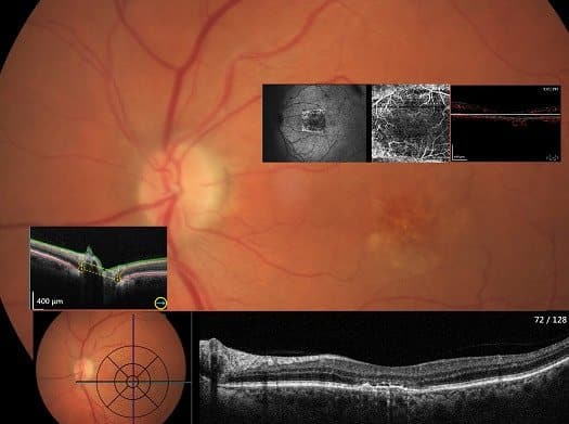
Peter D’Arcy investigates eye disease by a range of procedures including the latest canon ocular coherence tomography.
(OCT) scans determine if you have the earliest or advanced signs of macular degeneration
The OCT scans often employed in eye disease studies test for any distorted vision by the high resolution of retinal anatomy for abnormal blood vessels in the early stages of loss of central vision to legal blindness. It helps differentiate the two main types of AMD.
Macular degeneration types

OCT Angiography (OCTA) is a technology that reproduces retinal blood vessels by the reconstruction of retinal tomograms. With this technology, one can observe the retinal vascular state without fluorescein angiography, which is invasive and may cause a strong allergic reaction.
To generate the image of the retinal micro vascularisation, each B-scan of the examination pattern is consecutively
repeated several times. The contrast comparisons on consecutive B-scans at the same location show areas with a change in contrast over time and some areas where it remains constant. This is linked to the movement of red blood cells and hence the location of the vessels.
Amsler grid, Macular Scan,OCTA
Cover one eye at a time to look for any distortion evident on an Amsler Grid
Diabetic retinopathy and the neovascular type of age-related macular degeneration are the two most frequent retinal degenerative diseases causing the majority of blindness found in an eye exam.
Exudative retinal detachment can be caused by age-related macular degeneration, injury to the eye, tumours or inflammatory disorders


The OCT scans monitor for any distorted vision by the high resolution of retinal anatomy and can even be used as a sensitive early marker of dementia. OCT scanning may mirror changes going on in the blood vessels in the brain.
Optical coherence tomography (OCT) is a high-resolution, three-dimensional, noninvasive imaging technique. It is often called optical ultrasound as it is similar to ultrasound imaging but it obtains subsurface information from laser interferometry.

LAYERS OF POSTERIOR RETINA, CHOROID AND VITREOUS FLOATERS


Eye floaters may look to you like black or grey specks, strings, or cobwebs that move around when you move your eyes.
With age, the jelly-like substance (vitreous) inside your eyes becomes more liquid and the suspended fibres cast tiny shadows on your retina, called floaters.
The retina needs to be checked for any disturbance though.
READ MORE
The Centre for Eye Research Australia (CERA)has eg clinical trials for improving blood flow to the retina as there are many emerging treatments for geographic atrophy.
If you’re over 50 you are at higher risk of AMD and everyone with diabetes is at risk of vision loss through diabetic eye disease.
Blood vessels can be observed in a designated layer
Microaneurysm and non-perfusion areas can be identified.
CNV(Choroidal neovascularization) can be identified.
Blood vessels at the back of any leakage can be observed
Capillaries can be observed without using fluorescein
Frequent inspection is possible due to less burden on a patient with shorter inspection time
Blood flow visualization is possible in any depth from the retina to choroid however the actual leakage cannot be detected (requires fluorescein angiography.)
The vitreous is a transparent gel composed of water, collagen, and hyaluronic acid it occupies 80% of the volume of the eye, sometimes it can liquefy somewhat and cause floaters. Epiretinal membranes (ERMs) are sheet-like structures that develop on the inner surface of the neurosensory retina.
The macular changes that result from either ERM or VMT (vitro-macular traction) lead to similar symptoms: reduced visual acuity, metamorphopsia, difficulty using both eyes together, and even diplopia Epiretinal membranes may evolve between the neurosensory retina and a vitreous attachment. Vitrectomy does carry some risks (e.g., cataract, retinal tears, retinal detachment, endophthalmitis).
Regular eye examinations will ensure the best eye health possible. Stargardt’s disease is the most common form of juvenile macular degeneration. The second most common form of juvenile macular degeneration is Vitelliform macular dystrophy, also referred to as Best disease when it begins before age 6.
People with thinner retinas are more likely to have problems with memory and reasoning according to the dementia study JAMA
OCT scans have a role in risk profiling also for systemic stroke, myocardial infarction, hypertension, diabetes and an array of eye health disorders
Your eyes have aberrations overcome by their movements and have high resolution in a very small part of the eye responsible for the centre of your vision called the macula. You have a blind spot where your optic nerve takes the focused vision to the brain. To take in more information, you move your eyes around a scene to correct for these imperfections in your visual system.
MACULAR DEGENERATION TREATMENT
Eating for your eyes and healthy habits.
How many of these “healthy habits” can you tick off?
No smoking and a healthy, well-balanced source of antioxidants in foods and supplements.
Nuts, fresh fruit and vegetables
Fish two to three times a week
Low glycemic index (low GI) carbohydrates instead of high GI
Limiting intake of fats and oils
Maintaining a healthy weight,exercise regime and lifestyle.
Ensuring regular comprehensive eye tests including macula checks
The Omega 3 to Omega 6 ratio can be present in western diets 1:20 versus the healthier 1:4 in Mediterranean diets.
National Eye Institute USA Age-Related Eye Disease Study (AREDS) 2001 clinical trial showed high levels of antioxidants and zinc can reduce some people’s risk of developing advanced AMD by about 25 percent
Smoking harms lungs and your eyes. Smokers are four times more likely to develop macular degeneration than non-smokers.


The eyes require antioxidant activity to flush out toxins of oxidative stress produced as a result of metabolic reactions. Indeed certain patients can benefit from a specific mix of vitamins and minerals to slow the diseased condition. Every cell membrane has omega-3 fatty acids which has an anti-inflammatory effect compared with the Omega-6 type It is desirable to eat more Omega-3’s in our diet (oily fish, leafy greens etc.) Omega 6 plant oils needs to be reduced eg margarine, corn, sunflower, soybean in processed foods such as chips.
READ MORE
High levels of antioxidants and zinc significantly reduce the risk of advanced age-related macular degeneration (AMD) and its associated vision loss. These same nutrients had no significant effect on the development or progression of cataract. These findings from a nationwide clinical trial are reported in the October 2001 issue of Archives of Ophthalmology. The AREDS 1 formula is the first demonstrated treatment for people at high risk for developing advanced AMD.
The Areds2 study showed addition of lutein and zeaxanthin, DHA and EPA, or both to the AREDS formulation in primary analyses did not further reduce risk of progression to advanced AMD. However, because of potential increased incidence of lung cancer in former smokers, lutein and zeaxanthin could be an appropriate carotenoid substitute in the AREDS formulation.
Attention to diet eg particular fruit and vegetables eg being a good source of beta carotene (needed to create vitamin A )and antioxidants that also helps with the immune system as well as an array of medical approaches are required to limit potential vision loss. A diet rich in antioxidants involves:
The correct intake of plant based foods, carrots,eggs,milk and yogurt ,whole grains, quinoa, whole lentils, oats, brown rice ,oysters,red bell pepper,sunflower seeds, lentils, kidney beans, chickpeas, green peas, sprouts sweet potatoes among others are all vital for eye health as they supply the antioxidant activity.
Including the essential vitamins A,C,E omega-3,Zinc, Zeaxanthin,beta-carotene,lycopene,lutein,selenium.
Sources of antioxidants can be found in vitamins A, C and E, Dark chocolate, the minerals selenium, zinc and copper, and can also be found in phytochemicals from plants, fruits and vegetables. Too high doses of antioxidant supplements may work against your own antioxidants which are produced naturally rather than artificially.
Macutec once daily is an AREDS 2 formulation designed to reduce AMD implications from side effects of oxidative stress.
It is often recommended by your optometrist for those patients who prefer to take one capsule per day to reduce the risk of their dry age related macular degeneration progressing. AREDS-2 was an important five-year clinical study that demonstrated positive results for dietary lutein and zeaxanthin being a safe and effective alternative to beta-carotene.
It contains
Vitamin E 400 mg
Lutein 10 mg Zeaxanthin 2 mg
Zinc (from Zinc Oxide) daily dose of 25 mg
Copper (as Cupric Oxide) 2 mg
Ascorbic acid (Vitamin C) 500 mg
Omega-3 fatty acids – Eicosapentaenoic acid (EPA), and Docosahexaenoic acid (DHA).
The DHA in fish oil is essential for normal retinal functioning.
Macutec Once Daily doesn’t contain beta-carotene with its ill effects or fish oil unlike other alternatives eg Lacritec. Fish oil has a slight blood thinning effect so it is advised to consult your GP if you are taking blood thinning drugs such as warfarin.
Fish oil is an especially rich source of omega-3 fatty acids, which are also found in flaxseed, walnuts, and dark leafy greens.
There are no known significant drug interactions with Macutec Once Daily
The human body is capable of producing all the fatty acids it needs, except for two: linoleic acid (LA), an omega-6 fatty acid, and alpha-linolenic acid (ALA), an omega-3 fatty acid both found in plant and seed oils.
Omega-3s have anti-inflammatory benefits and help prevent heart disease, whereas Omega-6s lower blood cholesterol and support the skin.
Modern food processing and storage can rob many foods of much of their nutrition. For some natural sources it is difficult to actually eat the quantities required. Examples include fish and spinach. Even food preparation can reduce the nutrition content.
Macutec is based on a very large clinical trial which showed a significant benefit in slowing the progression of macular degeneration. The majority of the people who were on this trial and showed benefit were also eating healthily.
Sometimes Resveratrol in red wine is thought to help reduce abnormal angiogenesis.
AMD patients need to wear tinted glasses because of photostress. There can be many situations where the absorption is too dark or unsuitable ,requiring need for different filter values,sometimes more cheaply overcome by fitover shields of which we have in different densities. Photodynamic therapy (PDT) and the administration of compounds acting against vascular endothelial growth factor (anti-VEGF) are approved for the treatment of choroidal neovascularization (CNV) secondary to AMD.
Periodic intravitreal (into the eye) injections are sometimes required. Cell-penetrating peptide eye drops will drive the next generation of treatment for people with AMD rather than intraocular injections. Drugs that can activate proteins found in blood vessel cells are being developed to make the blood vessel more stable. Abnormal blood vessel growth and leakage are two primary factors in both age-related macular degeneration and diabetic retinopathy.
In the wet rather than the dry form of ARM anti vegf drugs some now plant based are normally administered by intravitreal injection rather than topical eye drops to reach the back part of the eye.
Photodynamic therapy uses laser light and light-sensitive dye to seal off the leaking areas. Free radicals, referred to as oxidants are unstable molecules in the body with unpaired electrons.
Oxidative stress plays a pivotal role in developing and accelerating retinal diseases including the leading cause of blindness in Austrlia -age related macular degeneration (AMD) the part of the eye responsible for central vision, glaucoma, diabetic retinopathy (DR), and retinal vein occlusion (RVO).
Risk factors for retinal vein occlusion which can cause other problems like heart attacks and strokes. The main risk factors are:
Age (most retinal vein occlusions happen in people over 60)
High blood pressure
High blood lipid levels
Diabetes
Smoking
Overweight
If a retinal vein occlusion causes fragile, abnormal new blood vessels to grow in the eye the risk of beeding can be minimised by using laser and/or intra-ocular drug injections.
If the central macula is affected and vision is reduced due to swelling of the central macula, treatment with anti-VEGF drugs may be used. Anti-VEGF drugs are administered as injections into the eye. The usual treatment regimen begins with monthly injections for three months. Then to maintain control of the disease, injections are typically continued on an indefinite basis, or until the swelling has resolved.
We also use digital retinal photography and employ tests to measure the effect on vision and if any distortion is present often involving an eye drop to dilate the pupil.
We can enhance vision by glasses,loupes and electronic magnification and with timely referral for laser intervention,injections or surgery.
Those with susceptible family history for ARM can consider predictive genetic testing, quit smoking, lower cholesterol, and be conscious of your environmental light exposure, and perhaps consider supplementation with antioxidants, such as lutein and zeaxanthin.
Blue light wavelengths boost our concentration so can impact on sleep
Blue light can damage the retina so at risk groups – family history of AMD, smokers, people who are obese,children can benefit by blue light filter especially when outside.
Blue light protection

Most AMD starts as the dry type and in 10-20% of individuals, it progresses to the wet type. You can have the early signs without knowing so intervention can be sought.
The Age-Related Eye Disease Study 2 (AREDS 2) was a multi-centre,randomised trial designed to determine the correct composition for macular supplements.
Macutec Essentials are the preferred option for people that need a smaller capsule option taken twice daily,otherwise Macutec Once Daily is recommended. Macular supplements contain just the right amounts of C and E vitamins along with zinc, lutein and zeazanthin to support your macular health.
AREDS antioxidant supplements remain the most proven way of slowing progression to late-stage AMD in high-risk patients.
MDeyes is Australia’s most affordable AREDS 2 capsule formula at $20.95 (RRP)
Experimental Eye Research, 79(6), 753-759, 2004
Macular Telangiectasia (“MacTel”)
Macular Telangiectasia (“MacTel”) is distinct from macular degeneration.
Type 1: congenital and unilateral
Type 2: acquired and bilateral. The most common form of the three types.
Type 3: Primarily occlusive phenomena which is quite rare.
With MacTel, the blood vessels around the fovea become dilated and leakage can be a sign of and cause of damage.
Type 1 MacTel involves localised dilations, or aneurysms and sometimes bleeding of blood vessels but without the “new” blood vessel growth characterised by widespread dilation and leakage
The MacTel Project is a collaboration involving more than 60 centres from around the world to better understand the progression of their disease


